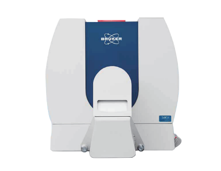
Bruker SkyScan Preclinical Micro-CT
Fastest scan time-3.9sec Scan time
Highest resolution-2.8μm Pixel size
Largest field of view-16 Mp CMOS Camera
Mouse and Rat Cassettes
Touchscreen Control
ON-SCREEN REAL-TIME DOSE METER
Integrated physiological monitoring
Detail
Product Principles
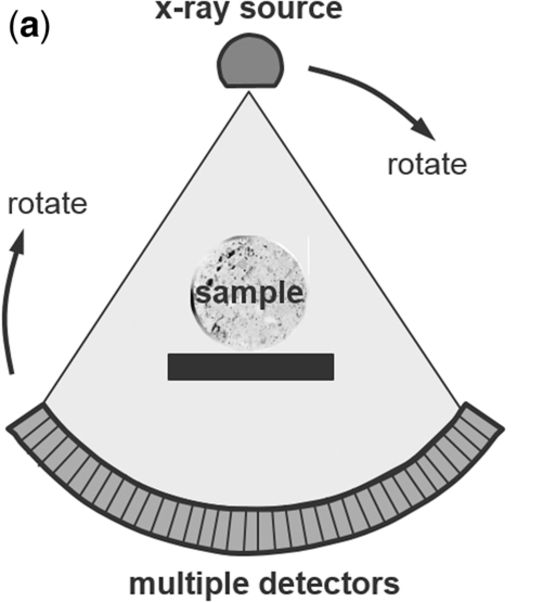
Micro-computed tomography (microCT) or X-Ray Microscopy (XRM) is X-ray imaging in 3D, the same method used in clinical CT scans, but on a small scale with massively increased resolution. It really represents 3D microscopy, where very fine scale internal structure of objects is imaged non-destructively, both in vivo and ex vivo. Bruker micro- and nano-tomography is available in a range of easy-to-use desktop and floor standing instruments, which generate 3D images of your sample’s morphology and internal microstructure with resolutions down to micron and even sub-micron level.
Brief Introduction
The SKYSCAN 1276 CMOS Edition is a high performance, stand-alone, fast, desktop in vivo microCT with continuously variable magnification for scanning small laboratory animals (mice, rats, ...) and biological samples. An optional Large Animal Transport System (LATS) is available which enables the system to mount also large animals including rabbits.
The SKYSCAN 1276 CMOS Edition has an unrivalled combination of high resolution, big image size, round and spiral (helical) scanning and reconstruction, and low dose imaging. The image field of view up to 75 mm wide and 310 mm long allows full body mouse and rat scanning. The variable magnification allows scanning bone and tissue samples with high spatial resolution down to 2.8 μm pixel size. Variable X-Ray energy combined with a range of filters ensures optimal image quality for diverse research applications from lung tissue to bone with metal implants. The system can perform scanning with continuous gantry rotation and in step-and-shoot mode with scanning cycles down to 3.9 sec. Furthermore, the SKYSCAN 1276 CMOS in vivo microCT administers a low radiation dose to the animals allowing multiple scans in longitudinal preclinical studies without the risk of unwanted radiation-induced side effects. The fully integrated physiological monitoring package allows monitoring and controlling the animal's wellbeing at all times through a video stream, ECG, temperature and breathing detection.
The SKYSCAN 1276 CMOS is complemented by 3D.SUITE. This extensive software suite covers GPU-accelerated reconstruction, 2D/ 3D morphological analysis, as well as surface and volume rendering visualization.
Product Characteristics
1. Mouse and Rat Cassettes
The SKYSCAN 1276 CMOS system is supplied with exchangeable animal cassettes that can be used in all Bruker in-vivo imaging instruments such as MRI, micro-PET, micro-SPECT, bio-luminescence, biofluorescence, etc. to collect multimodal information. It allows co-registration of functional and morphological information from the same animal.
2.Touchscreen Control
The user interface of the SKYSCAN 1276 CMOS system is simple and intuitive. The instrument can be controlled from the computer screen and also from the embedded force-sensitive touchscreen, which can be operated by gloved hands. The touchscreen allows selection of scanning protocol, adjusting the animal bed position and control of imaging and scanning. Where multiple scans are started from the touchscreen, the software will automatically save acquired data to separate subfolders with incrementally assigned folder names and dataset file prefixes.
3.ON-SCREEN REAL-TIME DOSE METER
The SkyScan1276 control software includes a real-time on-screen dose meter. It indicates an estimation of the dose absorbed by the animal body during scanning. The measurement is based on the absorption calculated from X-ray projection images of the animal cross-calibrated with electronic dosimeter measurements. The dose meter shows accumulated dose or dose rate. It is calibrated for X-ray absorption in the standard mouse and rat cassettes. In this way it measures the X-ray dose absorbed in animal body itself during scanning. The dose absorbed by the animal during a scan is documented in the scan log-file together with all scan and reconstruction settings.
4.Integrated physiological monitoring
The physiological monitoring system includes video monitoring of an animal with real-time movement detection, ECG and breathing detection, and temperature stabilization. A 5 megapixel color camera is mounted above the animal bed along with white LED illumination to introduce a real-time image of the animal during the scan. The software analyses the video stream from a user-selected area of the image where breathing movement is visible. These movements are converted into a movement waveform to provide breathing time marks for time-resolved reconstructions. The face mask on the animal bed is connected to an air/gas flow sensor for direct breathing detection. The ECG electrodes in the animal cassette are connected to a sensitive ECG amplifier. Both breathing and ECG signals are digitized and displayed as real-time profiles on-screen. The monitoring also includes temperature stabilization by heated airflow, which maintains the scanned animal at a selected temperature, to prevent cooling of the animal under anaesthesia.Application Fields
1. Mineralized Tissues
High resolution in vivo imaging of bone and teeth
True spatial resolution down to 6μm for visualizing the finest bone structures
Measure trabecular and cortical bone parameters
Analyze dynamic processes and skeletal disorders
2. Total body imaging
Body composition analysis for mice and rats
Contrast enhanced imaging of vascularisation, liver, spleen, etc
Tumor detection in various organs
2D kinetic study of bladder and kidney dynamics
3. Lung and Cardiac dynamic imaging
Prospective, retrospective and image based gating capabilities
Measure dynamic processes such as heart ejection fraction and respiratory volume
Lung tumour detection and analysis
Synchronized scans in only 2 minutes
4. Ex vivo sample analysis
Ex vivo scan capabilities with resolutions down to 2.8μm pixel size
Included sample holders for ex vivo sample analysis
Batch scan option for automated scanning of multiple samples
Application
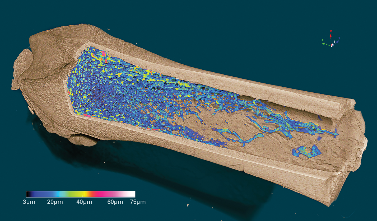
1. Volume rendered 3D model of a femur with color-codedrepresentation of the trabecular thickness, scanned at 2.8 umpixel size
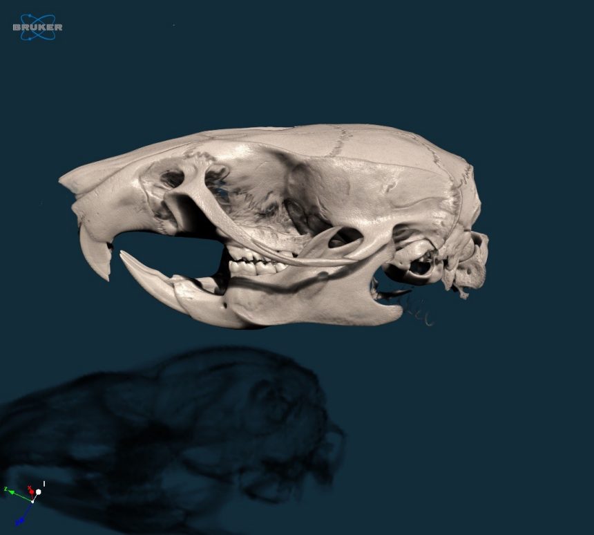
2. 3D model of a rat skull, scanned at 20um voxel size in vivo
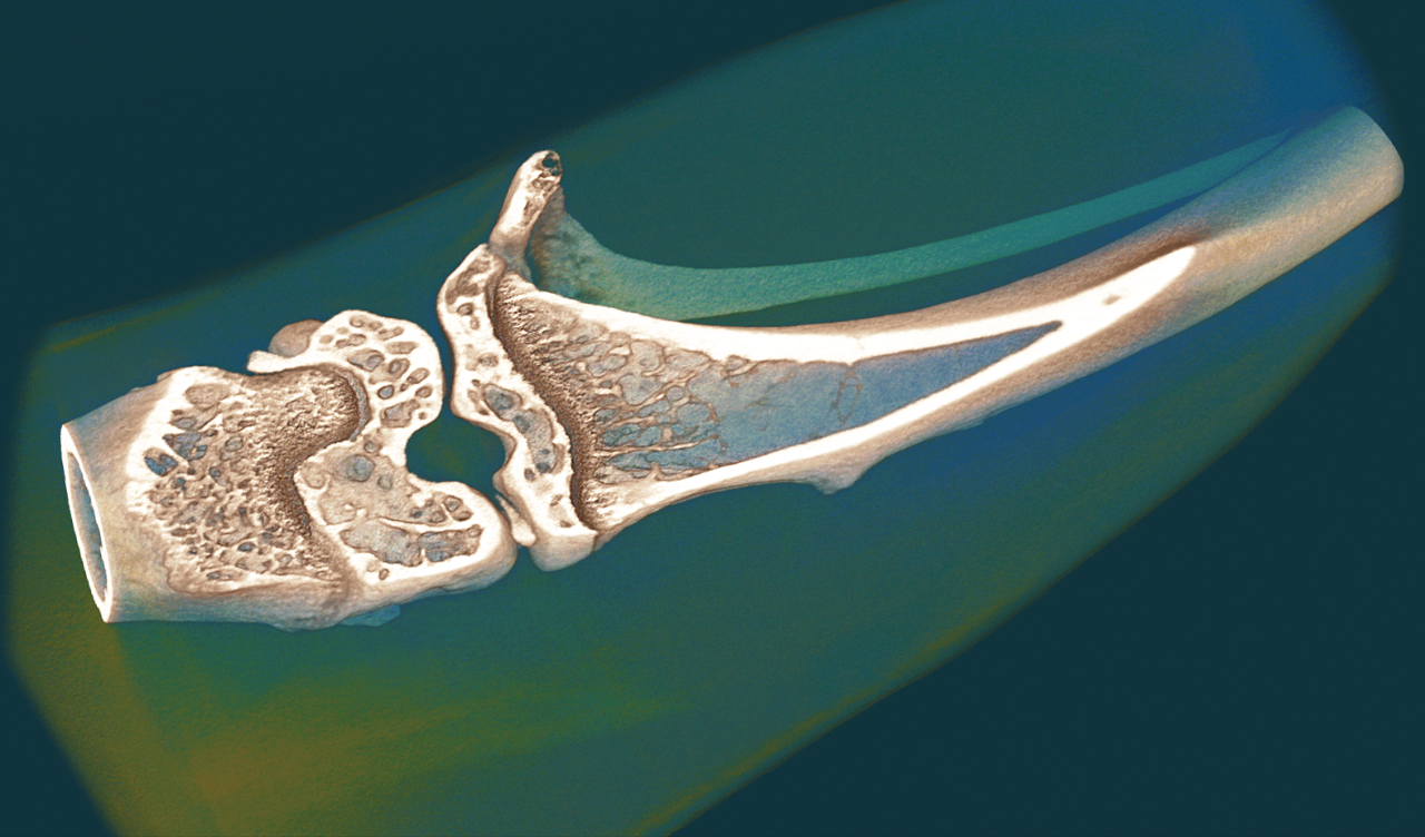
3. Volume rendered 3D model of a mouse knee, scanned at 6 umvoxel size in vivo.
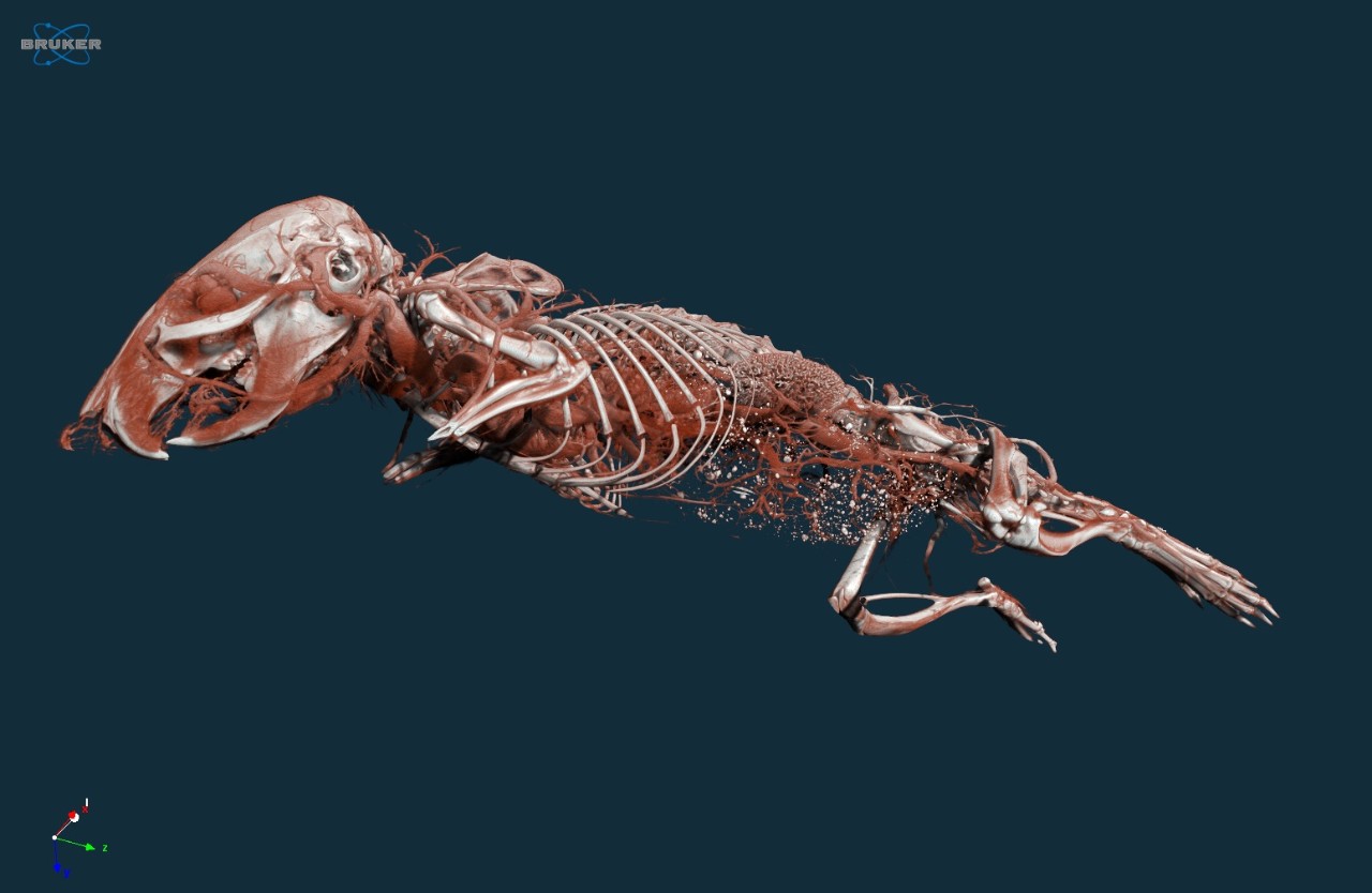
4. 3D representation of the mouse vasculature, scanned in vivoat 7 um voxel size after a bolus injection of vascular contrast agent.
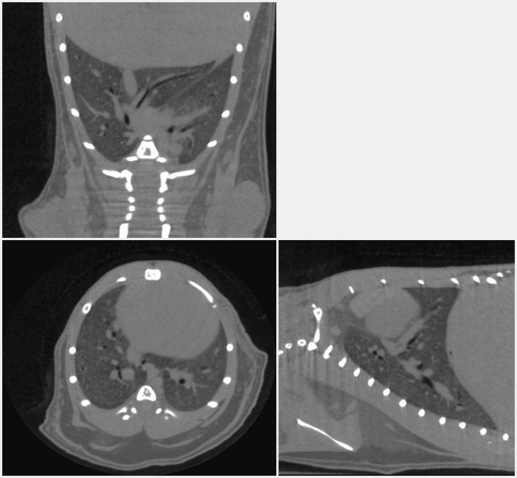
5. Orthogonal cross-sections through the mouse lung scannedin vivo, showing the blood vessels and large airways inside the lung.
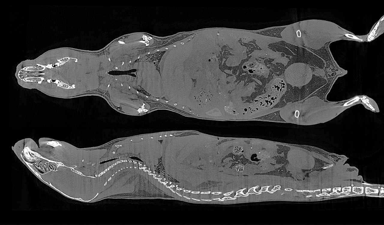
6. Cross-sectional slices through a mouse body, scanned at 17um voxel size in vivo without contrast agent injection.
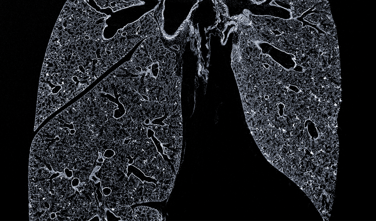
7. Cross-section through a mouse lung, scanned ex vivo afterchemical drying at 3 um voxel size.
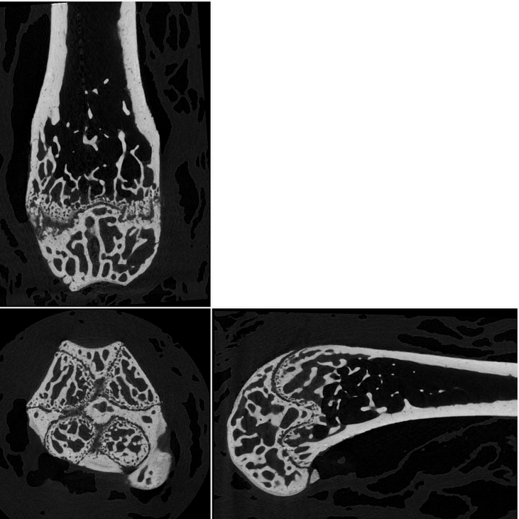
8. Orthogonal slices through a mouse femur, scanned at 2.8 umvoxel size.
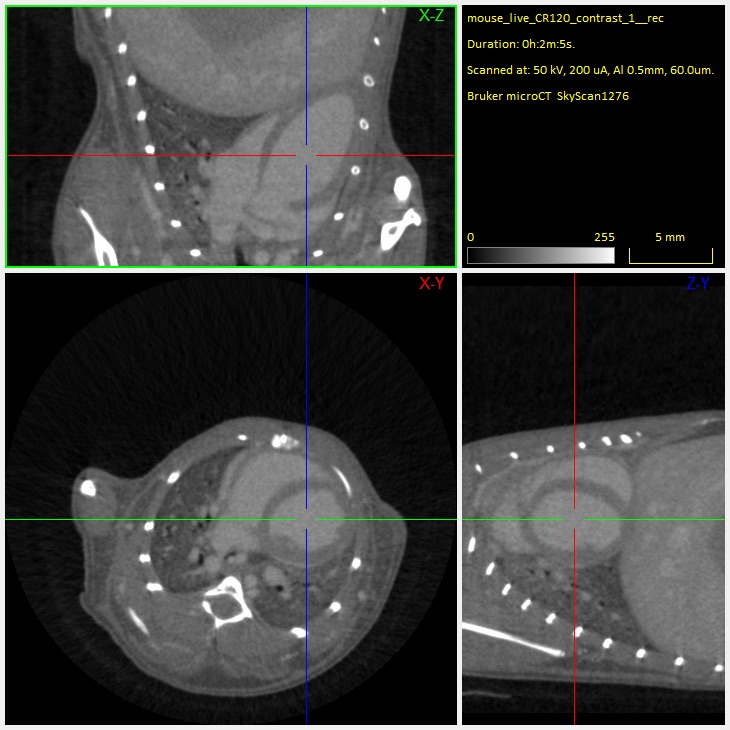
9. Orthogonal slices through a mouse heart, scanned in vivoafter contrast agent injection.
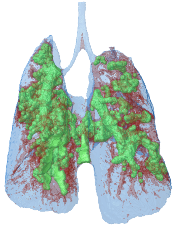
10. 3D model of a mouse lung, made transparent to visualize thelung tumour tissue in green.
